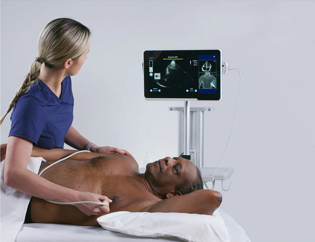
A cardiac ultrasound also called echocardiography or an echocardiogram is an examination of the heart. This is done by a medical specialist called a cardiologist. This procedure utilizes sound waves to produce images of the heart’s four chambers and valves. It allows your doctor to detect any abnormalities.
Cardiac ultrasound can be used to detect heart disease or injury. Also, to diagnose congenital or perforated defects. Additionally, it is used to evaluate valvular disease, measure blood flow across the valve (called regurgitation). Another use case is to assess ventricular hypertrophy and other problems relating to areas outside the valves; they can also rule out other possible ailments such as an aortic aneurysm or pulmonary embolism.
Cardiac ultrasound is, therefore, a non-invasive, rapid, inexpensive medical imaging technique that allows the visualization of structures and blood flow in the heart. There are multiple types of echocardiography that use different scanning techniques: visualizing blood flow throughout the body by point-by-point; real-time echocardiogram; three-dimensional echocardiography or a Triple-Headed Color Doppler Ultrasound Scanner: such as the Triple-Headed Color Doppler Ultrasound Scanner: Vsono-CL1
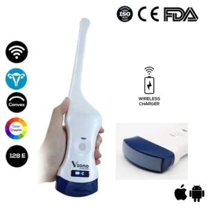
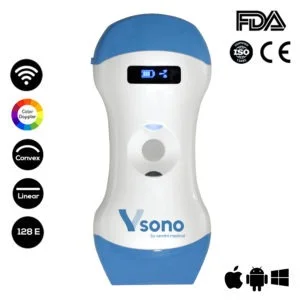
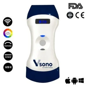
The Vsono-CL1 has 3 probes in 1 device: A convex, linear, and cardiac (phased) array. The phased array of the Vsono-CL1 sends high-frequency sound waves into the tissues and organs, causing the visible movement of a specialized gel medium.
The Color Doppler feature allows the visualization of blood flows from and out of the heart.
“near real-time information about the direction and character of intracardiac flows. Color Doppler is especially useful in the diagnosis and quantitation of valvular regurgitation, and in the detection of intracardiac shunts”.
(Perry & Nada, 1988)
The results obtained by echocardiography can help to evaluate patients with congestive heart failure. Also, monitor a patient during or after surgery, or assist with diagnosis in cases of congenital heart disease.
References:
Herbst et al. (2021)
Aly et al. (2021)