Using ultrasound in the assessment of a woman’s ovaries searching for polycystic ovary syndrome has become an essential test. In fact, ultrasonography helps To achieve a successful diagnosis of the condition. Polycystic ovary syndrome is a hormonal disorder that affects approximately 8-12% of women under the age of 40 PCOS is caused by an imbalance in the hormones progesterone and insulin.
What can cause this hormone imbalance? For some women, stress or weight gain may lower the levels of progesterone. Obese or women who have high levels of sugar in their blood often deal with insulin resistance down the line. And finally, genetics may play a role too.
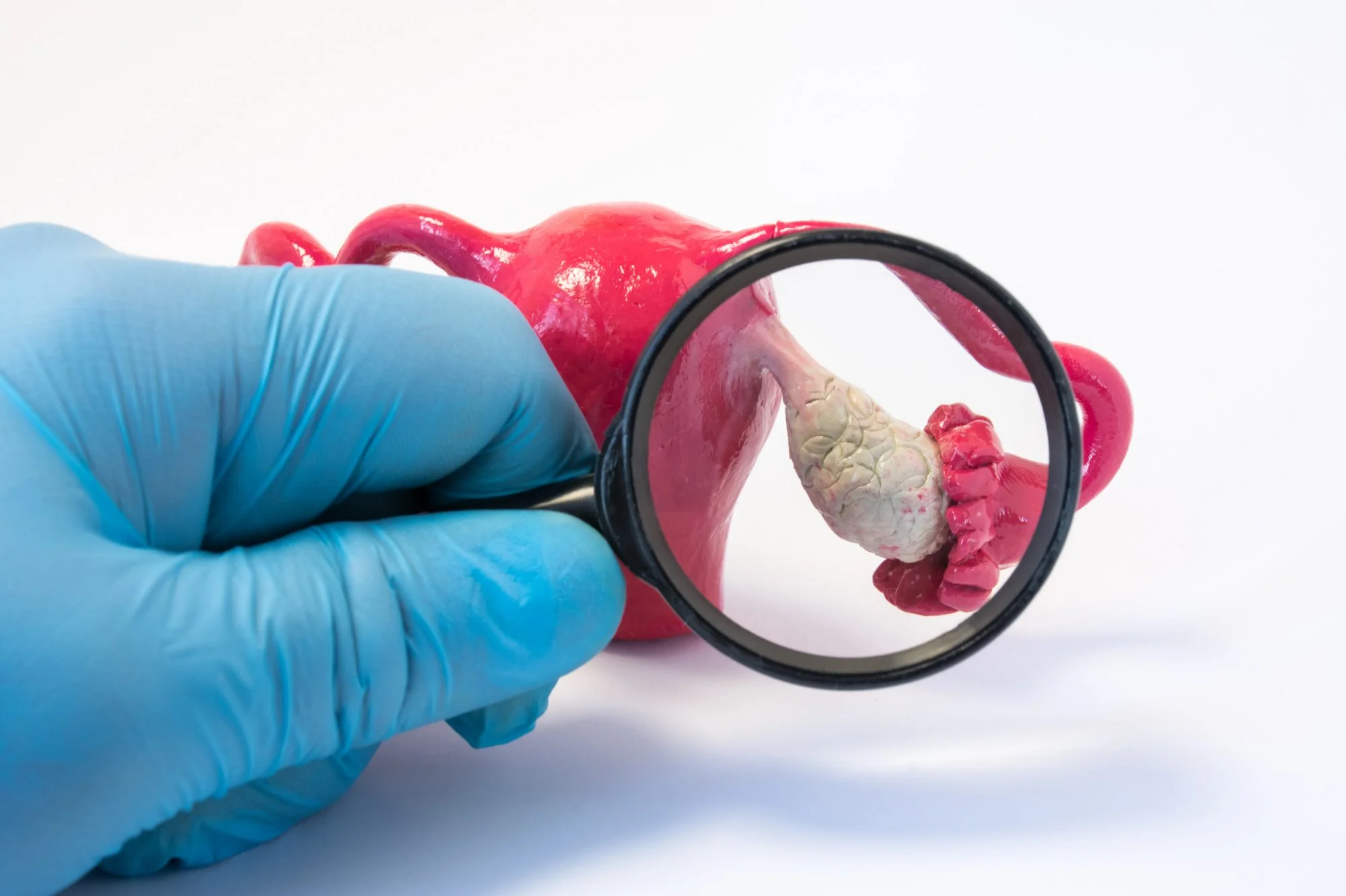
The OB/GYN will perform an ultrasound if their patient exhibits signs of polycystic ovary syndrome, such as irregular periods. A patient with the syndrome may have cysts on her ovaries. Enlarged follicles on her ovaries due to high levels of male hormones called androgens are also among the main symptoms.
If a woman is experiencing irregular menstrual cycles, obesity, infertility, acne, or excessive hair growth on the face and body then it’s likely that she has the syndrome.
Polycystic ovary syndrome ultrasound scanning is a clinical test for women with irregular or no periods. Ultrasound is a painless and non-invasive procedure that involves inserting a probe into the vaginal canal. It usually takes three minutes or less. The probe sends sound waves into body tissues. This creates echoes they decode as pictures of the organs inside your body.
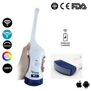
The Color Doppler Transvaginal Ultrasound Scanner: Vsono-TVU1 is quite useful in this procedure. The Vsono-TVU1 contains 2 probes in 1 device: A transvaginal probe + A convex probe, allowing for a thorough and detailed scanning.
References:
PCOS