


Transvaginal Ultrasound: A Complete Guide to Uses, Procedure & Safety

Everything you need to know about endovaginal ultrasound exams for pelvic health and pregnancy.
What Is a Transvaginal Ultrasound?
A transvaginal ultrasound (also known as endovaginal ultrasound) is a diagnostic imaging exam that uses high-frequency sound waves to produce detailed images of the female reproductive organs, including the uterus, ovaries, cervix, fallopian tubes, and vagina.
Unlike abdominal ultrasound, where the probe is placed on the skin, this exam involves gently inserting a thin, lubricated probe about 2–3 inches into the vaginal canal. Because the probe is closer to the pelvic organs, the images are much clearer and more precise.
When Is It Recommended?
Doctors may suggest a transvaginal ultrasound to:
-
🔍 Investigate pelvic pain or abnormal bleeding
-
🌡️ Evaluate infertility causes
-
🤰 Monitor early pregnancy (heartbeat, ectopic pregnancy, miscarriage evaluation)
-
🩺 Check IUD placement, detect ovarian cysts, fibroids, or pelvic infections
-
👁️ Assess cervical changes linked to preterm labor risk
How to Prepare
-
Bladder prep – In some cases, a full bladder is required. Drink about 32 oz (1 liter) of water one hour before, if advised by your doctor.
-
No tampons – Remove tampons if menstruating.
-
Clothing – Wear easily removable lower garments; you will change into a gown.
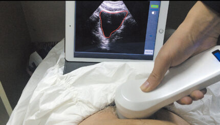
What to Expect During the Exam
-
You’ll lie on an exam table with feet in stirrups, knees bent.
-
A lubricated, condom-covered probe is gently inserted into the vagina.
-
The probe emits sound waves to generate real-time images of the pelvic organs.
-
The technician may rotate the probe slightly for different angles.
⏱️ The test usually takes 15–30 minutes.
💡 Discomfort is typically mild, similar to a Pap smear. If you have a latex allergy, request a non-latex cover.
Advanced Option: Saline-Infused Sonography (SIS)
For a more detailed uterine evaluation, sterile saline can be introduced into the uterus to expand the cavity and highlight abnormalities.
⚠️ Not performed during pregnancy or in case of active pelvic infection.
Safety of Transvaginal Ultrasound
-
✅ No radiation exposure – relies only on sound waves
-
✅ Safe during pregnancy – no proven harm to mother or baby
-
✅ Minimal discomfort – pain is rare, but notify your provider if you experience severe discomfort
Conditions That Can Be Detected
-
Ectopic pregnancy
-
Ovarian cysts / uterine fibroids
-
Placenta previa
-
Pelvic infections (inflammation, abscess)
-
Reproductive cancers (screening for ovarian or uterine tumors)
After the Procedure
-
Results – If your doctor performs the scan, results are often available the same day. If reviewed by a radiologist, expect 24–48 hours.
-
Follow-up – A transabdominal ultrasound may be suggested if additional views are needed.
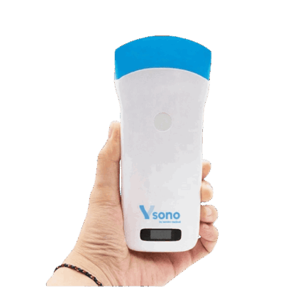
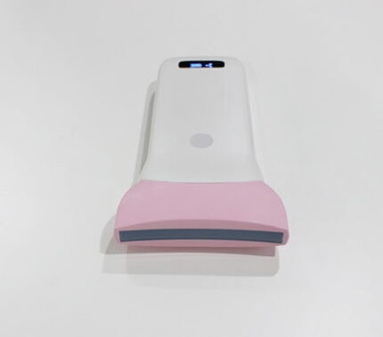
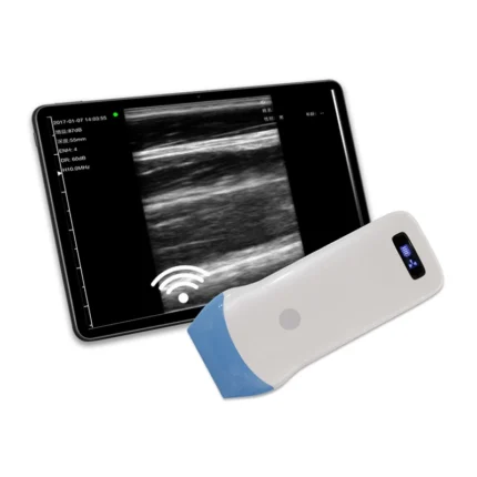
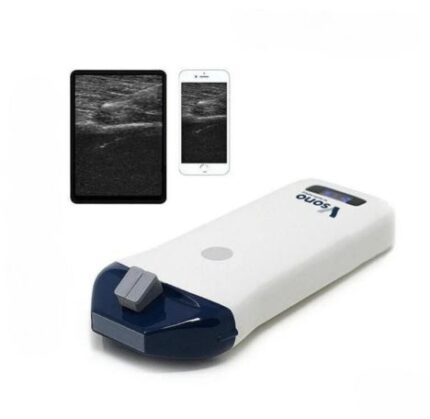
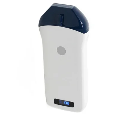
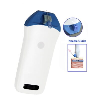
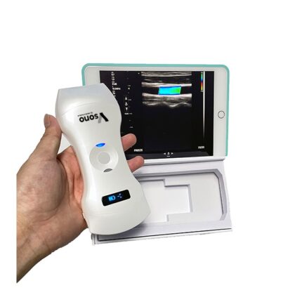
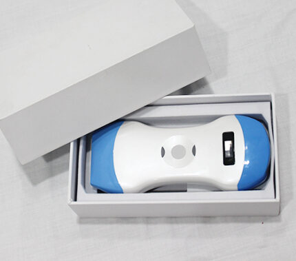
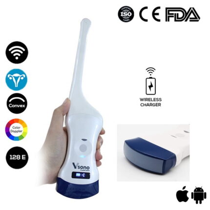
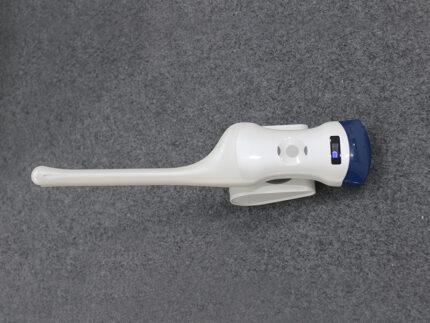
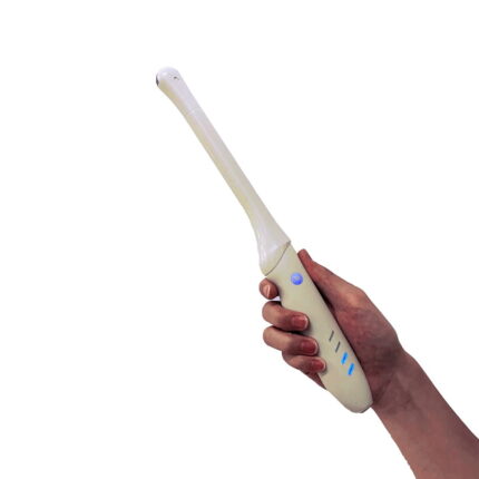
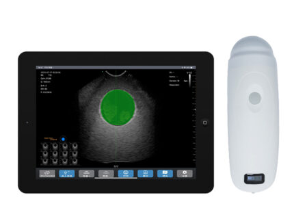
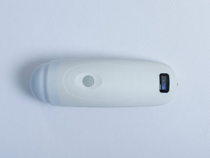
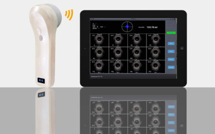
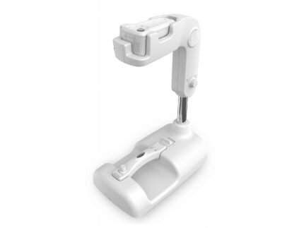
Featured Device: Color Doppler Transvaginal Ultrasound Scanner — Vsono-TVU1
To highlight how this exam is performed with advanced medical technology, here is a professional-grade device available at Vendra Medical:
👉 Discover the Color Doppler Transvaginal Ultrasound Scanner Vsono-TVU1
This scanner combines two probes in one (convex + transvaginal) and offers wireless operation, making it ideal for modern clinical practice.
Conclusion
A transvaginal ultrasound is a safe, reliable, and essential diagnostic tool in gynecology and obstetrics. With its ability to deliver clear, high-resolution images, it plays a key role in detecting conditions early and supporting effective treatment. The use of advanced equipment such as the Vsono-TVU1 further enhances diagnostic accuracy and patient comfort.