


Enhancing Thyroid Diagnostics: The Role of High-Resolution Point-of-Care Ultrasound
Thyroid Diagnostics are essential when dealing with disorders that represent a significant portion of endocrine pathologies encountered in clinical practice. While initial assessment relies on thyroid function tests (primarily TSH levels), high-resolution ultrasonography remains the gold-standard imaging modality for the morphological evaluation of the thyroid gland. Its non-invasive nature, lack of ionizing radiation, and superior spatial resolution for superficial structures make it indispensable. This tool is crucial for general practitioners, endocrinologists, and radiologists alike in their thyroid diagnostics efforts.
The Diagnostic Pathway: From TSH to Imaging
The clinical approach to a suspected thyroid disorder is well-established. The process begins with serological testing of Thyroid-Stimulating Hormone (TSH), with normal values typically ranging from 0.5 to 5.0 mIU/L. Thyroid diagnostics do not stop there; imaging plays a key role.
When TSH levels are abnormal, or when a physical examination reveals a palpable nodule or goiter, further investigation is warranted. Ultrasonography is the critical next step to characterize the gland’s structure and identify abnormalities. It guides subsequent management decisions, such as the need for a fine-needle aspiration (FNA) biopsy, showcasing its vital role in thyroid diagnostics.
Key Clinical Applications for Thyroid Ultrasound
High-resolution ultrasound provides crucial information across a spectrum of thyroid conditions, a core component of thyroid diagnostics.
- Nodule Characterization: It is the most sensitive method for detecting and characterizing thyroid nodules. Key features such as echogenicity, composition (solid vs. cystic), margins, shape (taller-than-wide), and the presence of microcalcifications are evaluated. These features help to stratify the risk of malignancy, often using a system like TI-RADS.
- Diffuse Thyroid Disease: In autoimmune conditions like Hashimoto’s thyroiditis or Graves’ disease, ultrasound can reveal characteristic parenchymal changes. These include diffuse hypoechogenicity or a heterogeneous echotexture.
- Guiding Interventional Procedures: Ultrasound guidance is standard practice for FNA. It ensures accurate needle placement within a target nodule and increases diagnostic yield while minimizing sampling error.
- Monitoring and Surveillance: It is essential for monitoring the size and characteristics of known nodules over time. It is also vital for post-operative surveillance in patients with thyroid cancer, cementing its place in ongoing thyroid diagnostics.
Essential Technology for Optimal Thyroid Imaging
Performing a high-quality thyroid examination requires specific ultrasound technology to ensure diagnostic accuracy, forming an integral part of thyroid diagnostics.
- High-Frequency Linear Transducer: A high-frequency probe, typically in the 7.5 MHz to 10 MHz range or higher, is mandatory. This provides the necessary axial resolution to visualize fine details of the thyroid parenchyma and the subtle characteristics of nodules.
- Color Doppler Imaging: This feature is not merely an adjunct but a necessity. Evaluating vascularity within and around a nodule is a key component of risk stratification. Furthermore, Color Doppler is instrumental in identifying the “thyroid inferno” pattern characteristic of Graves’ disease. It is also crucial for assessing inflammatory states in thyroiditis.
The Vsono-L1: A Solution for Point-of-Care Thyroid Assessment
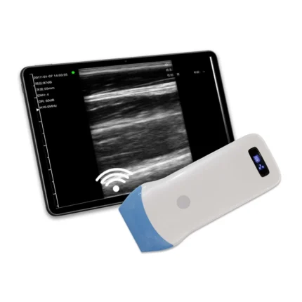
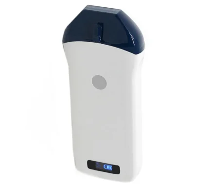
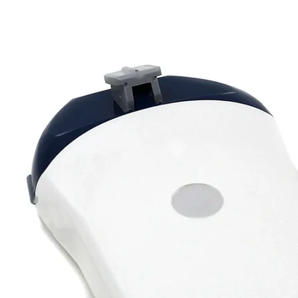
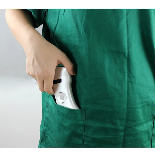
For clinicians seeking to integrate high-resolution imaging into their practice efficiently, the Vendra Medical Vsono-L1 Wireless Linear Ultrasound Scanner presents a compelling solution.
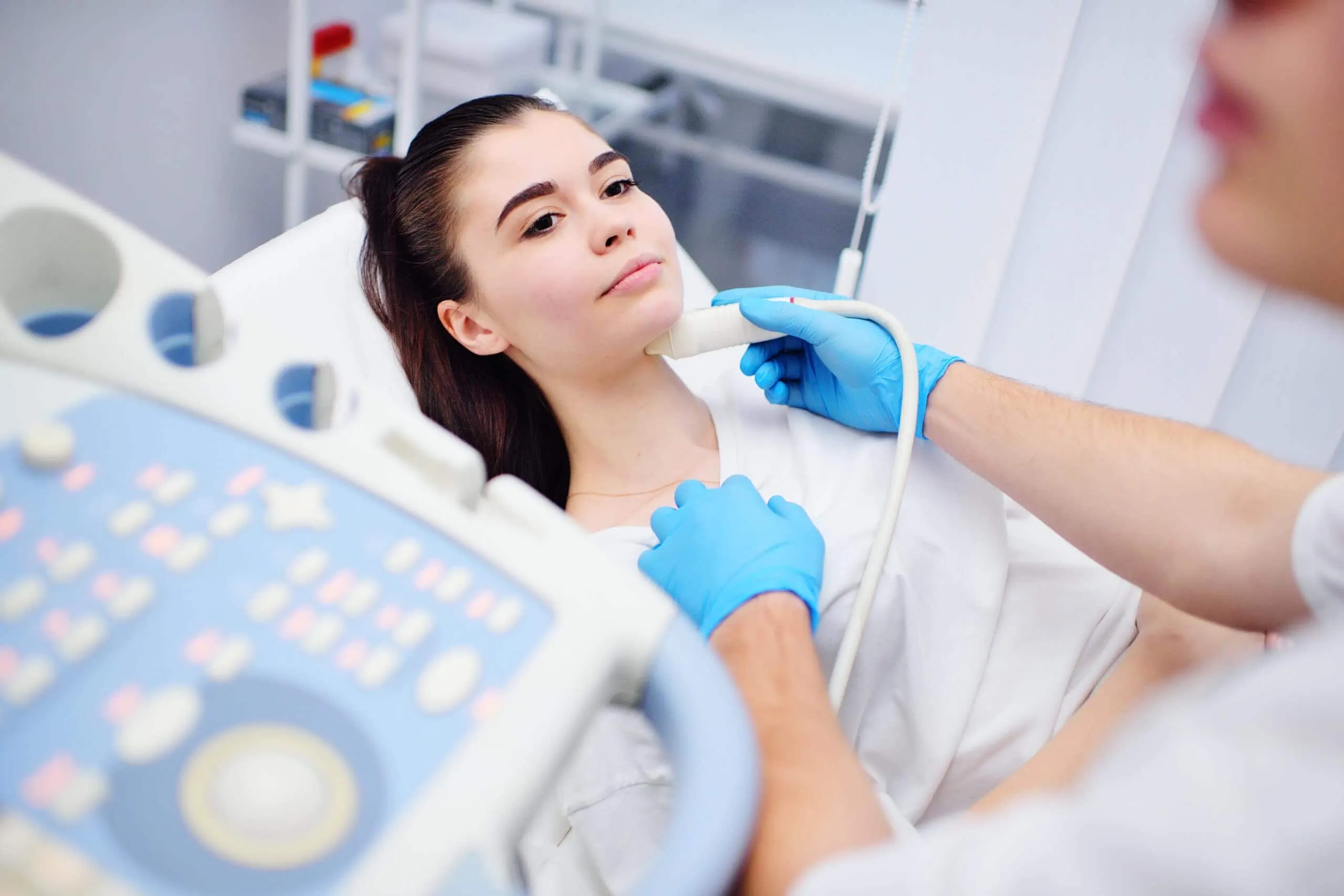
The Vsono-L1 is a portable, battery-powered device designed for point-of-care (POCUS) applications. Its wireless design eliminates the constraints of traditional cart-based systems. This makes it ideal for use in outpatient clinics, hospital wards, or for guiding procedures directly in the examination room.
Crucially, the probe’s frequency range of 7.5 to 10 MHz aligns perfectly with the requirements for high-resolution thyroid imaging. This allows the Vsono-L1 to deliver clear, detailed superficial scanning. It facilitates accurate diagnosis and informed clinical decision-making right at the patient’s side as part of the comprehensive thyroid diagnostics array of tools.
